Leticia Gallardo Estrella, Lead Deep Learning Engineer at Thirona, shares the recent developments in vascular phenotyping of pulmonary disease, elaborating on the possibilities and benefits of AI-powered pulmonary vessel differentiation.
AI may be a buzzword in many industries, but for us, the medical industry, research community and clinical professionals, its potential impact on patient care and scientific progress is undeniable. With the power to speed up diagnoses and identify new therapeutic targets or interactions, AI is a tool that can help us save more lives and push the boundaries of medical innovation.
Pulmonary blood vessels
Pulmonary blood vessels are vital elements of our respiratory system, carrying oxygen-rich (pulmonary veins) and oxygen-poor (pulmonary arteries) blood from and to every inch and corner of our lungs.
As veins and arteries in the lungs serve different, yet critical purposes in the body, their altered morphology can be symptomatic for certain pulmonary vascular diseases (PVDs). These diseases might manifest as, e.g., blocked, narrowed, or enlarged veins or arteries. Importantly, the type of vessel affected can point at the exact cause of the patient’s problems. Diagnosing PVD can be difficult, frequently being quite intrusive and the differences between arteries and veins are nuanced and not easy to detect. This leads to delays in detection or misdiagnosis, missed treatment opportunities and worse patient outcomes.
Recent advancements in medical research propose that accurate distinction and quantification of these blood vessels in diagnostic methods will be a major step forward in diagnosing pulmonary vascular diseases. AI unlocks this possibility, introducing exemplary precision in detection of vascular changes in patients’ lungs by analyzing high-resolution computed tomography (HRCT) scans.
With this blog, I want to elaborate on the possibilities and benefits of AI-powered pulmonary vessel differentiation. This article will give you insights into technological progress in pulmonary vascular diseases detection with the use of AI-powered algorithm. I will also present the prospects of pulmonary vessels phenotyping in diagnostics, research and clinical trials based on the first results of the studies conducted with our new pulmonary biomarkers.
Let’s begin with the outlook on pulmonary vascular diseases and their detection.
Artery/vein phenotyping in pulmonary vascular patients
Know your enemy
Recent developments in diagnostics of PVDs suggest that differentiation between veins and arteries is vital to accurately detect the root of the problem [1, 2].
The most prominent example of a misdiagnosed PVD is pulmonary hypertension (PH) that can affect veins or arteries (or both). Each type of pulmonary hypertension has different causes which determines the approach to treatment [3].
However, current diagnostic tools lack proper artery/vein (AV) phenotyping, which can be instrumental in understanding vascular changes occuring in PH.
Diagnosing pulmonary vascular diseases today
The main diagnostic tools to detect pulmonary vascular diseases include right heart catheterization (RHC), electrocardiography (ECG), thoracic computed tomography scan (CT), and multiple lung function tests. These methods have their limitations and are not always enough to provide a clear understanding of the patient’s condition.
RHC is the gold standard for diagnosis and classification of PH, but it is also the most invasive of the diagnostic methods for PVDs. Even though it is commonly used to diagnose PH, physicians are often challenged with the interpretation of unclear results. Studies show that this method, while necessary to confirm PH, is often performed without a standardized protocol which causes misdiagnoses [5].
Echocardiography can inform us about the heart condition, but it has low specificity. Right heart problems observed on echocardiography might suggest numerous disorders: pulmonary hypertension, arterial embolism, or Chronic Obstructive Pulmonary Disease (COPD), among others. While it's valuable information, it does not point to the exact cause of the patient's struggles and requires further investigation into the etiology of their symptoms.
CT is an imaging modality with great potential in diagnostics the evaluation of the pulmonary vasculature [4]. However, the challenge lies in the analysis of the scans. Manual analysis of the CT results is time-consuming and isn’t practical for clinical settings, especially large-scale clinical studies. To facilitate diagnostic efforts, a patient is often given intravenous iodinated contrast. This radiocontrast agent, while effectively visualizing the vasculature, may have adverse effects for the patients.
Automatic pulmonary AV phenotyping in non-contrast CT scans will help overcoming these issues, facilitating an accurate characterization of vascular changes in a non-invasive way.
But how can it actually help patients? And what impact could it have on our research?
Artery/vein differentiation in diagnostics and research
The principal reason for pulmonary AV differentiation is to uncover new insights into pulmonary vascular disease diagnosis, underlying disease mechanisms, and treatment response.
Emerging studies show that precise phenotyping of the pulmonary vasculature can help identify specific disease subtypes, potentially leading to more informed treatment decisions and more effective patient monitoring.
Our expectation is that AV phenotyping will have the most noteworthy benefits in detecting pulmonary hypertension and its subtypes.
Pulmonary (arterial) hypertension identification
Pulmonary hypertension is characterized by high blood pressure in pulmonary vasculature. Although many PH patients show similar initial symptoms, this disease has been divided into five subtypes varying in their primary causes. Depending on the PH cause and whether veins or arteries (or both) are affected, patients require different treatment.
Although PH has no cure, there are various promising treatment options available nowadays. For PH patients it’s especially important to choose the right treatment path in the early stage of the disease to delay or prevent its progression.
The problem with early diagnosis lies in the lack of specific symptoms. However, the primary studies using AI-powered LungQ™ AVX module (Thirona) show that early-stage pulmonary hypertension can be detected in thoracic CT scans. PH is a rare disease with a very high underdiagnosis rate [6]. Applying AI to help with its detection can potentially result in slowing the disease progression in PH patients by ensuring they get the best possible treatment sooner.
Additionally, the subtype-specific symptoms are not easily detectable. As an example, PH group 1 (PAH) and PH group 3, while of different etiopathology, present similar symptoms and hemodynamic profiles. To ensure proper diagnosis and cause detection, patients are burdened with redirection to specialized medical centers for further investigation. They may need to undergo further tests which involve invasive procedures and come at an additional cost.
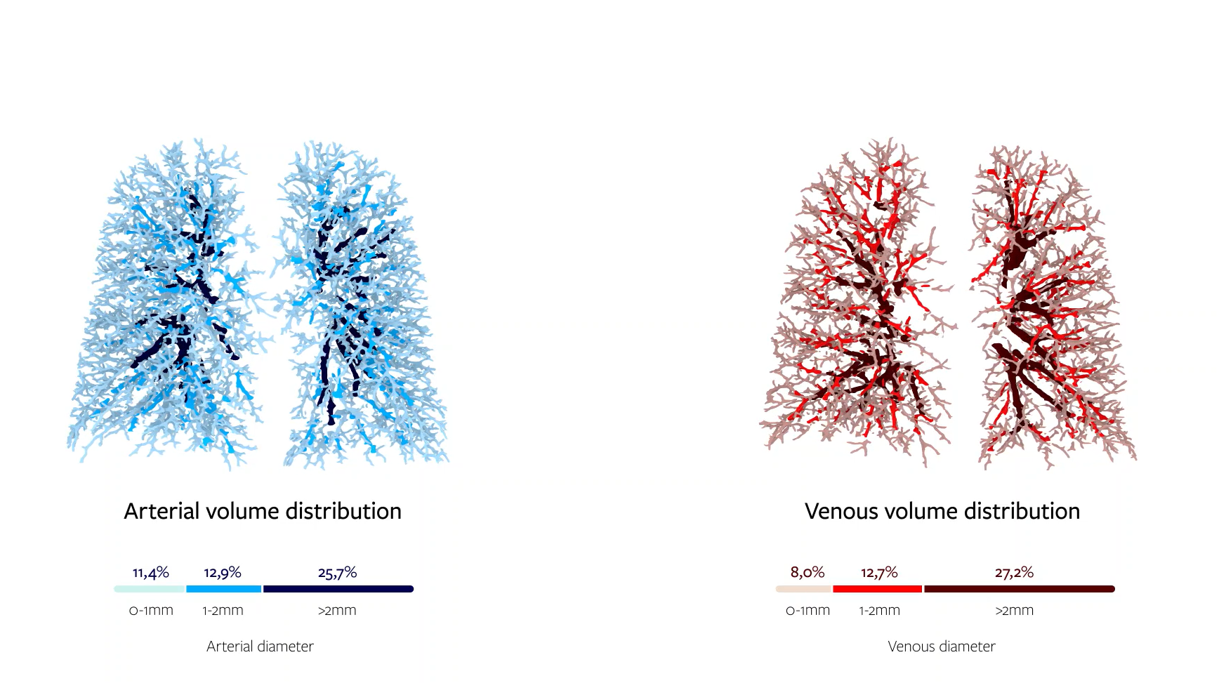
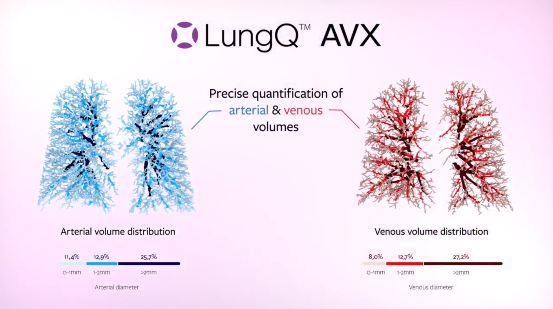
This could soon change with the adoption of AI-assisted diagnostics. Studies show that LungQ™ AVX algorithms not only can detect PH, but also identify different subtypes of this condition. The example from our study in collaboration with the University Hospital of Liege [7] suggests that PAH patients exhibit significant differences in the overall arterial and venous volumes compared to patients with PH group 3. This is a big step forward in the distinction between these two diseases.
But P(A)H is not the only condition that can be aided with the automatic AV phenotyping.
Pulmonary vascular disease exploration
When thinking about the potential applications of AV phenotyping, we can’t forget about novel research. The newly unlocked potential of non-invasive distinction between pulmonary arteries and veins opens the possibility of searching for new dependencies between various factors and symptoms. This will increase our knowledge with valuable insights of disease progression and development. Moreover, with the high throughput ensured by automation, it can help conduct large-scale clinical studies in PVDs which require observation of patient outcomes. With the use of AI, these observations become more accurate and faster than ever.
We already know of many lung diseases with vascular issues, yet the extent of these vascular components and their details are often poorly understood. This can be said about COPD or Cystic Fibrosis, topics of ongoing research. AV phenotyping might provide us with exploration potential leading to development of new treatments and better understanding of these diseases.
The power of AI in pulmonary artery/vein phenotyping
As AI adoption increases, the rapid and accurate analysis of vast amounts of CT scan data is starting to transform disease diagnostics, treatment planning and development of new therapies. In the field of pulmonary vascular diseases, AI, and in particular artery/vein phenotyping, has the potential to revolutionize the ways we treat these conditions.
However, the use of AI always raises a lot of questions about the inner workings of the algorithm.
LungQ™ AVX: wealth of data

Concluding 8 years of research and continuous innovation efforts by Thirona’s Deep Learning team, we have just introduced a new automatic AI-based algorithm called AVX. It performs deep characterization of the pulmonary vasculature in diseases such as pulmonary hypertension. AVX is a valuable addition to our AI-based lung quantification platform LungQ™ platform, allowing for computed AV phenotyping analysis. This new feature of LungQ™ follows many previously validated and certified solutions for quantitative assessments of various lung biomarkers on chest HRCT scans.
So how does it work?
To put it simply, LungQ™ AVX processes thoracic non-contrast HRCT scans, segmenting the arteries and veins visible in the scan. The software is able to accurately quantify vessel diameters and volumes down to the subvoxel range, which is crucial to obtain a precise picture and ensure that no changes go unnoticed.
The quantified volume of the patient’s pulmonary arteries and veins can facilitate accurate phenotyping, and can serve as a diagnostic support, but also to measure treatment outcomes. Additionally, AVX quantitative characterization of pulmonary arteries and veins can enable faster and more accurate medical and pharmaceutical research.
Benefits of artery/vein phenotyping
This novel measurement capability of LungQ™ AVX provides insights that could previously only be qualitatively estimated from a CT scan. Enabling detailed quantifications throughout the entire vascular tree, it opens new perspectives for treatment of pulmonary hypertension.
AVX's precision in classifying pulmonary vascular abnormalities and characterizing vessels up to the subvoxel range is vital to achieving early diagnosis in terms of pulmonary hypertension, since it has the potential to detect vascular alterations even before they are picked up by current global hemodynamic measurements such as pulmonary artery pressure or pulmonary vascular resistance.
LungQ™ AVX does not require using contrast for the HRCT scan to look deep into patients’ pulmonary vasculature. Its sensitivity and accuracy allow for quantification of the blood vessel volumes in routine CT scans without any unnecessary inconvenience for the patients.
All these advantages potentially mean new and better treatments that can be developed, better diagnostic solutions and the elimination of diagnostic delays or misdiagnosis. Ultimately, it would allow us to attain precision in diagnosis and more effective, personalized treatments.
If you’re representing the medical industry, working on new treatments for PH diseases, don’t hesitate to connect with us to discover how we can partner to help you innovate faster in this field.

